Neuroimaging
(brain scans)
(brain scans)


| Neuroimaging
techniques provide another tool for diagnosing dementia-related diseases in
living subjects. |
|
| Various forms of
equipment have been developed, all of which rely upon the properties of
chemistry and physics to reproduce two-dimensional images of brain
structures. Within the field of
neuropsychiatry, radioactive (CT, PET,
and SPECT) and non-radioactive (MRI, MRA) methods are commonly employed in
order to track changes in the appearance of the brain. |
|
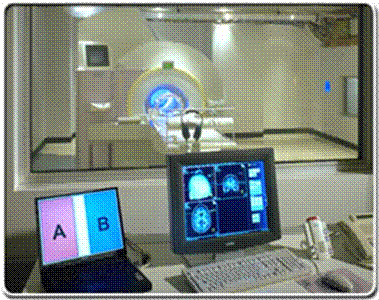
| This picture
shows an MRI machine (Magnetic Resonance Imaging). |
|
| Using MRI
technology, scientists are able to discern the features of different
components within the brain (e.g., white matter vs. gray matter, fluid vs.
solid tissue). The goal of anatomical
studies is to take “snapshots” of the brain in order to identify normal or
abnormal structures. So-called
“functional” brain scans (fMRI) compare changes in blood flow which accompany
specific activities or states of mentation. |
|